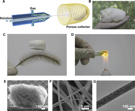
Fig. 1. Synthesis and structural characterization of TiO2 nanofiber sponge.

(A) Schematic of a solution blow-spinning. (B) Photograph of a macro-sized Ti(OBu)4/PVP precursor sponge. (C) Ultralight TiO2 sponge standing on a setaria viridis. (D) Sponge heated by an alcohol lamp without damage, indicicating good heat resistance. (E) SEM image of millimeter-sized TiO2 sponge. (F) Zoomed-in section of TiO2 sponge. The image shows the cellular fibrous structure and the uniform distribution of nanofibers. (G) Transmission electron microscopy (TEM) image of a TiO2 nanofiber.
