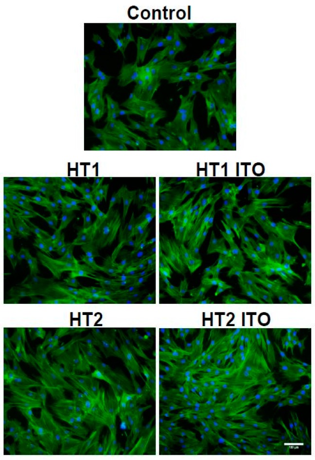
Figure 8.
Actin cytoskeleton organization of dermal fibroblast cells after 4 h of incubation with cotton knit treated with TiO2-1% Fe-N samples. F-actin (green) was labeled with phalloidin-fluorescein isothiocyanate (FITC), and nuclei (blue) were counterstained with 4′,6-diamidino-2-phenylindole dihydrochloride (DAPI). Scale bar: 100 µm.

