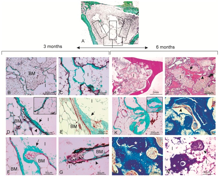
Figure 3.
Histological analysis of the material samples stained with Masson’s Trichome (A–G,K–N) and hematoxylin-eosin (H,J) three months (A–G) and six months (H–N) after implantation. (A) panoramic image of the implant section together with the different areas drawn for the descriptive histological study as being representative of the histological study at three and six months. The ceramic implant, which was removed during histological processing, is seen as a blank/empty space; (B,C) the whole material is observed in close relation with the hematopoietic bone marrow-interior of the pores colonized by hematopoietic bone marrow elements (BM: bone marrow; I: implant); (D) blood capillaries located between a thin osteoid tissue layer and surrounding bone marrow material. The insert of the figure shows some areas of the new osteoid tissue with osteocytic lacunae (osteoblast) and with osteocytes inside (BM: bone marrow; I: implant, arrow head: capillary blood; arrow: osteoid); (E) image corresponding to the central area of the material with newly formed bone tissue with the trabecular bone characteristic (arrow); (F,G) new bone colonization of the implanted material’s interior (arrow); (H) after six months of implantation, the implanted material is completely enveloped by new bone tissue and penetrates large areas (* cortical bone); (J) Presence of bone tissue in both the periphery and interior of the implanted material; (K) bone tissue deposition occurred mainly in the form of layers. The insert shows a pore filled with new bone formation; (L) bone with normal mature characteristics and in-growth inside one of the implanted material’s pores; (M) differences in the blue staining shade reveals the presence of scattered areas of older bone tissue (light blue) (arrow) and newly formed bone (dark blue) (*); and (N) presence of two nodular structures within the material. Cuboidal osteoblastic cells are seen to line the surface of the developing osteoid tissue (arrow) (BM: bone marrow; I: implant).

