Figure 3.
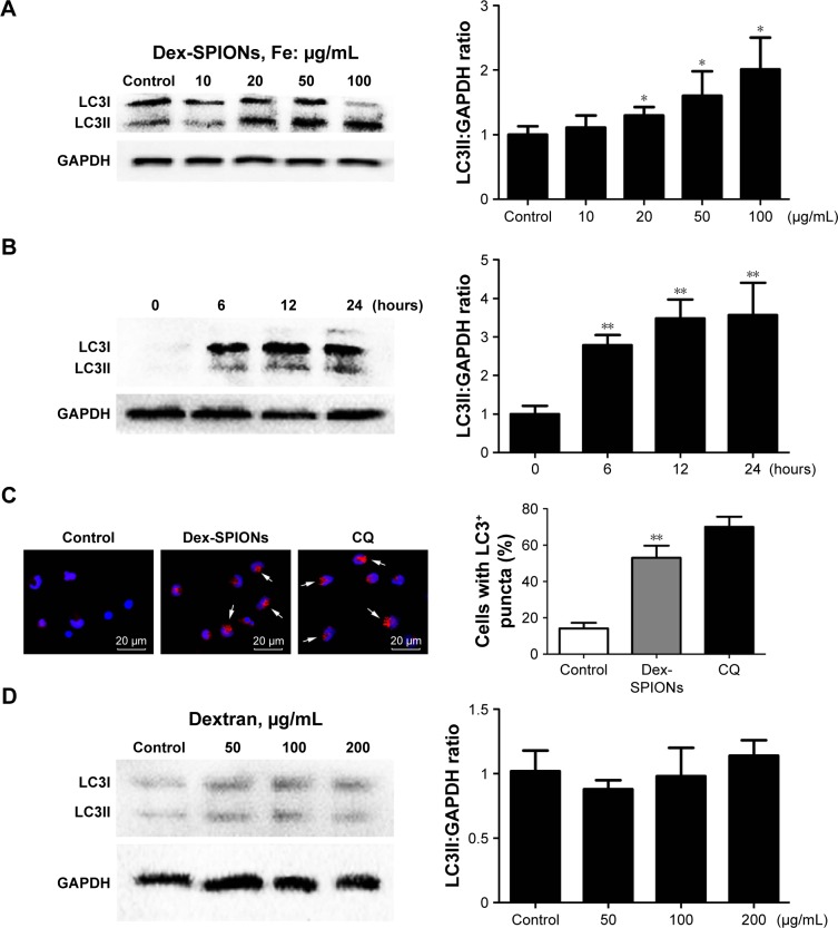
Dex-SPIONs induced autophagosome accumulation in human monocytes.
Notes: *P<0.05; **P<0.01; n=3. (A) Western blot analysis of LC3 in human monocytes after treatment with various concentrations (10–100 μg/mL) of Dex-SPIONs for 24 hours. (B) Western blot detected LC3 in human monocytes after treatment with Dex-SPIONs (100 μg/mL) at the times indicated. (C) Human monocytes treated or not with Dex-SPIONs (100 μg/mL) for 24 hours, stained with anti-LC3, DAPI (blue), and Alexa Fluor 568 goat antimouse IgG (red), then processed for confocal microscopy. CQ (30 μM) was used as a positive control. Quantitation of the percentage of cells with LC3+ puncta (white arrows). Scale bars 20 μm. Magnification ×60. (D) Western blot analysis of LC3 in human monocytes after treatment with various concentrations of dextran for 24 hours.
Abbreviations: Dex-SPIONs, dextran-coated superparamagnetic iron oxide nanoparticles; DAPI, 4′,6-diamidino-2-phenylindole; CQ, chloroquine.

