
Fig. 3. Structure of TPR2 TPD in complex with D53 EAR-2 peptide.
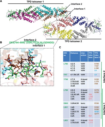
(A) Structure overview showing two TPD tetramers with an EAR-2 peptide at the TPD tetramer-tetramer interface (dashed rectangle). EAR motif peptides are shown as green stick models, the TPDs are shown as cartoon models, and the TPD Zn2+ ions are shown as gray spheres. (B) Close-up of the interface; key amino acids and bonds are shown and labeled. D794 and Q804 of the EAR-2 motif (letters in parentheses) were not resolved in the structure. (C) Main interacting residues between TPR2 TPD and the D53 EAR-2 motif peptide. Brown, TPD monomer 1; cyan, TPD monomer 2; pink, TPD monomer from interacting tetramer [same color code in (A)]; bold, key interaction residues. aa, amino acids; VdW, van der Waals bond (4.5 Å cutoff); H-bond, hydrogen bond (3.5 Å cutoff).