
Fig. 2. Localized VEGF enhances vascularization in extrahepatic transplant sites.
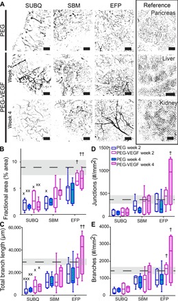
(A) Recipients of PEG-only or VEGF-presenting hydrogels were lectin-perfused at 2 or 4 weeks, and excised grafts were whole mount–imaged. Scale bars, 200 μm. Vascular characteristics of blood vessel fractional area (B), total branch length (C), junction number (D), and branch number per field of view (FOV) (E). Dashed line and shaded region represent average and SEM for pancreas reference, respectively. Minimum to maximum box-and-whisker plots, n = 5 to 7 per group. † versus SUBQ within the same time point (††P < 0.01 and †P < 0.05); × versus pancreas control (×××P < 0.001, ××P < 0.01, and ×P < 0.05); evaluated by Kruskal-Wallis nonparametric tests with Dunn’s multiple comparison.