
Fig. 5. In vivo bioluminescent islet tracking allows real-time monitoring of islet survival.
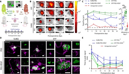
(A) Gels containing Luc+GFP+/B6 hybrid islet grafts, either with or without VEGF, were delivered to extrahepatic sites as demonstrated in the schematic. (B) Representative in vivo bioluminescence images. (C) Recipients were monitored weekly for bioluminescent signal (left) by intraperitoneal luciferin injection, and cumulative bioluminescent data after day 7 demonstrate significantly enhanced survival in EFP-VEGF group. (D) Lectin perfusion at experimental end point allowed visualization of Luc+GFP+/B6 islet graft [GFP (green)] integration with host vasculature [lectin (magenta)]. High-magnification images of EFP/PEG and EFP/PEG-VEGF islets illustrate integration of vasculature with islet organoid structure. (E) Time-to-peak bioluminescent signal serves as an additional measure of graft vascularization over time. Error bars represent SEM. n = 3 to 4 per group. ****P < 0.0001, ***P < 0.005, and **P < 0.01, evaluated by Kruskal-Wallis nonparametric tests with Dunn’s multiple comparison. †P < 0.05 versus SUBQ-PEG, evaluated by one-way ANOVA with repeated measures and Dunnett’s multiple comparisons test. Scale bars, 100 μm.