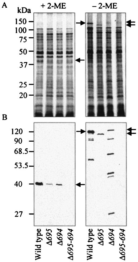
FIG. 2.
Typical protein patterns and Western blots of envelope fractions. Bacterial envelopes were denatured in SDS with or without 2-ME at 100°C for 5 min and then loaded onto SDS-polyacrylamide gels. The gels were stained with CBB (A) and subjected to Western blot analysis with anti-Pgm6/7 serum (B). Each 50-μg protein was applied to each lane of the gel. Arrows show the predicted monomers and trimers that consisted of Pg0695 (Pgm6 monomer, 41 kDa) and/or Pg0694 (Pgm7 monomer, 40 kDa). Monomers of Pgm6 and Pgm7 were not discriminated from each other in SDS-PAGE assays.

