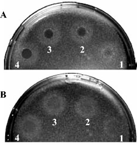
FIG. 10.
Lysozyme sensitivity of sigV mutant strain S26 (A) compared to that of the wild-type JH2-2 strain (B). A dilution of 106 cells of each strain ml−1 taken at 1 h after the onset of stationary phase was plated on LB agar to give confluent colonies. Immediately after plating, 10 μl of egg white lysozyme at 40 (spot 1), 60 (spot 2), 80 (spot 3), or 100 (spot 4) mg ml−1 was spotted onto the plates. Zones of clearing (dark circles) were photographed after 24 h of incubation at 37°C.

