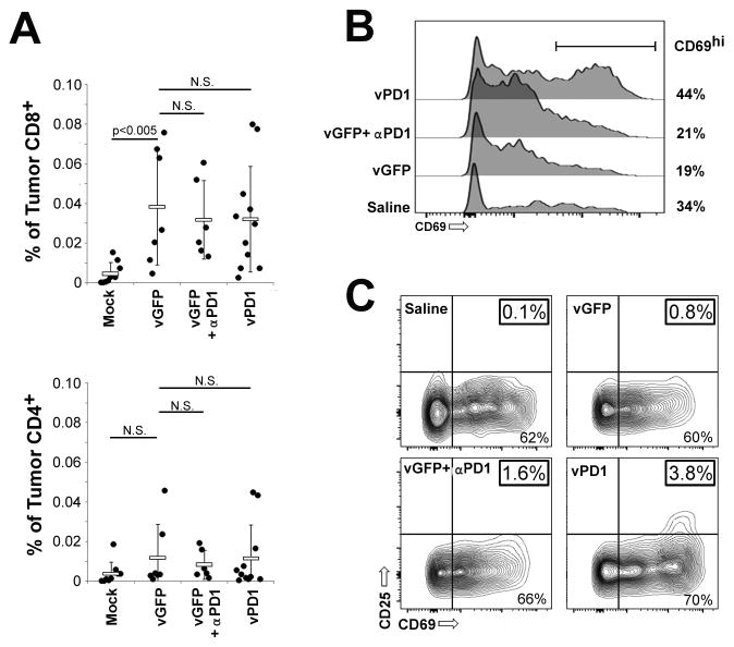
Figure 6. vPD1 enhances activation of CD8+ TILs.
(A–C) C57/B6 mice were injected SQ with 4x10⁵ B16/F10 cells. On day 7, mice were randomly separated into one of four cohorts: saline (n=9), vGFP + isotype (n=7), vGFP + αPD1 antibody (n=6), or vPD1 (n=11). Viral injections consisted of 1x10⁷ FFU and were given on days 7, 9, and 11. Antibody injections consisted of 100μg/injection and were given on days 7 and 11. On day 12, mice were euthanized, tumors disassociated into single cell suspensions, and TIL’s extracted using Histopaque. TILs were then analyzed using flowcytometry. (A) Numbers of CD4+ and CD8+ cells as percentage of total viable cells. (B) Surface expression of the early activation marker CD69 on CD8+ TILs. (C) Surface expression of the early and late activation markers CD69 and CD25 on the surface of CD8+ TILS. Data represents summation of four independent experiments. Significance was determined using unpaired students T-Test.

