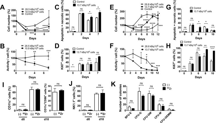
Figure 2. 89Zr-oxine-labeled BM cells retain differentiation function.

A. BM cells were labeled with 89Zr at 10.9 (8.14–13.7, filled circle) and 28.5 (28.1–28.8, cross) kBq/106 cells and cultured with SCF, FLT3-L and TPO (100 ng/ml each, n=6). The cells labeled at the lower dose slightly proliferated after day 4, while the cells labeled at the higher dose decreased in number (10.9 kBq/106 cells vs control (open circle): P = 0.0305, 0.0015 and 0.0001, 28.1 kBq/106 cells vs control: P = 0.0068, 0.0002, and 0.00001, at 2, 4, and 7 days, respectively). B. Decay corrected specific activity of the 89Zr-oxine labeled BM cells decreased in the cells labeled with the lower dose, reflecting the cellular proliferation, but remained constant in cells labeled with the higher dose (n=6) (P = 0.00197, 0.00035 at day 4 and 7, respectively). C. Higher fractions of annexin V+ and/or PI+ cells were detected by flow cytometry in the 89Zr-labeled cells (13.7 kBq/106 cells, black, n=3) on day 4 and 7 than the non-labeled cells (white). D. The labeled cells proliferated as indicated by Ki67 staining (n=3). E. BM cells were labeled with 89Zr at 13.7 (filled circle) or 28.8 (cross) kBq/106 cells and cultured with 20 ng/ml GM-CSF (n=6). Compared to the non-labeled control (open circle), the labeled cells showed delayed increase in cell number (13.7 kBq/106 cellls: p<0.0001 on day 6 and 8, p=0.0168 on day 12, 28.8 kBq.106 cells: p=0.0121 and 0.0001 on day 3 and 12, p<0.0001 on day 6, 8 and 10, vs control). F. Decay corrected cell associated 89Zr activity gradually decreased in cells labeled at the lower dose, but was plateaued with the higher dose until around day 8 (p=0.0177, 0.0008 and 0.0006 on day 8, 10, 12, 13.7 vs 28.8 kBq/106 cells). G. Higher fraction of annexin V+ and/or PI+ cells were detected in the 89Zr-labeled cells (13.7 kBq/106 cells, black) on day 6 and in the non-labeled cells (white) on day 8 (p=0.0176 on day 6 and 8). H. Proliferation of the cells indicated by the expression of Ki67 was delayed in the labeled cells reaching the peak on day 8, whereas the peak in non-labled cells was observed on day 6 (p=0.0041 and p<0.0001 on day 8 and 10). I. 89Zr-oxine-labeled BM cells (13.7–22.2 kBq/106 cells, black) differentiated to DCs (CD11c+) comparable to the non-labeled cells (white) after a 10 day-culture in GM-CSF (20 ng/ml). Around 40% of CD11c+ cells expressed a maturation marker CD86 (n=6). J. When cultured with IL-15 (25 nM), 89Zr-labeled BM cells (black) differentiated to NK1.1+ NK/NK-T cells in 10 days similar to the non-labeled cells (white) (n=6). K. Colony forming cell assay of non-labeled (white) and labeled (14.8 kBq/106 cells, black) BM cells showed no significant difference in the number of various hematopoietic cell colonies on day10 (n=3, BFU-E: burst forming unit-erythroid, CFU-G: colony forming unit-granulocyte, CFU-GM: colony forming unit-granulocyte macrophage, CFU-M: colony forming unit-macrophage, CFU-GEMM; Colony forming unit-granulocyte, erythrocyte, macrophage, megakaryocyte).
