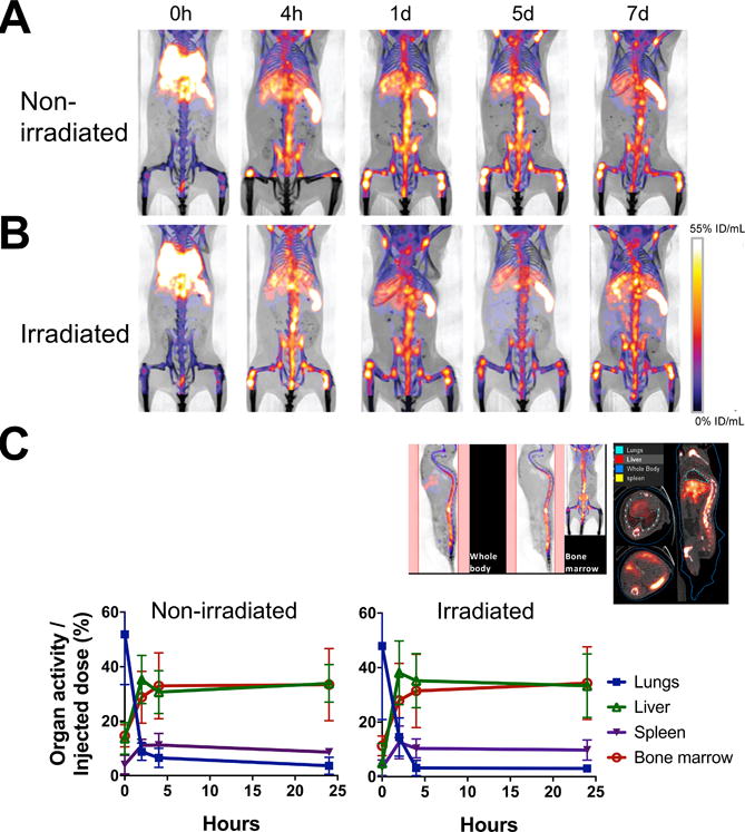
Figure 3. Serial microPET/CT imaging reveals rapid trafficking of 89Zr-oxine-labeled BM cells through the lungs to the BM, spleen and liver.

A. 89Zr-oxine-labeled BM cells (2×10⁷ cells at 16.6 kBq/106 cells) were transferred via the tail vein to non-BM ablated (A) and BM ablated (9.5 Gy whole-body irradiation) (B) mice (n=5). PET images indicate that immediately after the injection (0 h), cells started to migrate to the lungs, liver, spleen and the BM (%ID/mL: percent of injected dose per mL of tissue). C. Quantitative analysis of the microPET/CT images revealed similar migration kinetics to the BM, lungs, liver and spleen between non-irradiated and irradiated recipient mice (n=3, blue squire: lungs, green triangle: liver, purple triangle: spleen, red circle: bone marrow). The example of quantitated area/regions of interest set on the images are indicated in the upper left (bone marrow) and right (lungs, liver, and spleen).
