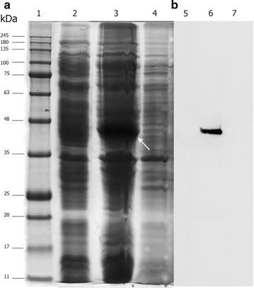
Fig. 2.

The expression analysis of anti-CD22 scFv–apoptin in the soluble and insoluble fractions of lysed bacterial cells. a SDS-PAGE analysis: protein molecular weight standards (lane 1), total protein from E. coli BL2 (DE3) containing pET-28 a (+) vector (negative control) grown under identical condition (lane 2), insoluble fraction (lane 3), and supernatant of cell lysate (lane 4) of induced E. coli BL2 (DE3) containing pET-anti-CD22 scFv–apoptin. A major band of ~44 kDa in insoluble fraction indicates anti-CD22 scFv–apoptin (white arrow). b Western blot analysis: the samples were loaded as in a
