
Figure 2.
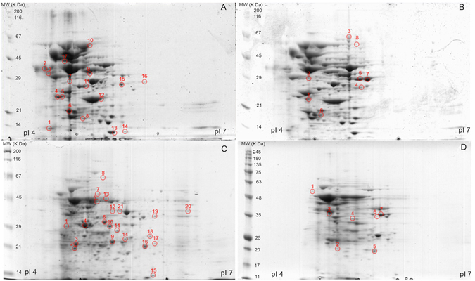
Representative 2-D gels of cellular proteins of S. pneumoniae TIGR4 (A and B) and R6 (C and D) cultured without (A and C) and with 0.5 × MIC purified rhodomyrtone (B and D). The isolated proteins were separated by isoelectric focusing in the pI range of 4 to 7 in the first dimension (11 cm). The proteins were further separated by 10% SDS-PAGE in the second dimension. Spot numbers indicate spots with altered abundances: those marked in (A and C) diminished after rhodomyrtone treatment, and those marked in (B and D) augmented after rhodomyrtone exposure.