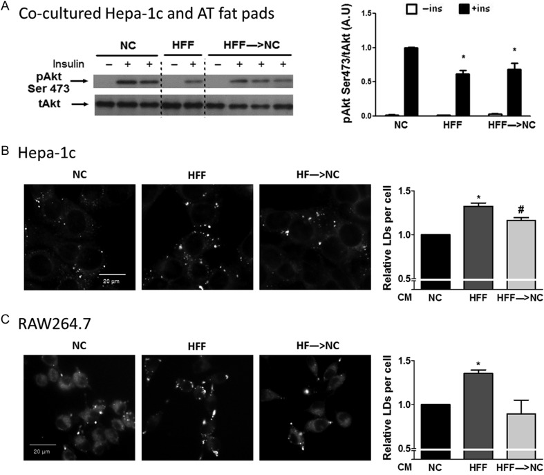
Figure 5.
Functional reversal of increased liver cell and macrophage lipid storage by adipose tissue conditioned medium. (A) Insulin-stimulated Akt phosphorylation in Hepa-1c hepatoma cells exposed to conditioned media of adipose tissue from NC, HFF, and HFF→NC mice. Shown is a representative blot and densitometry analyses normalized to the signal in insulin stimulated cells pre-treated with conditioned media of NC mice. N = 4–20 individual wells from 2 independent experiments, *P < 0.05 vs NC. Hepa-1c hepatoma cells (B) and RAW264.7 macrophages (C) were treated with adipose tissue conditioned media (AT-CM) for 6 h. Lipid droplets (LDs) were stained with BODIPY 493/503 neutral lipid dye and imaged by Operetta high content imaging system. Graphs represent the relative number of LDs per cell, compared to NC treated cells. The results are expressed as mean ± s.e.m. of five (Hepa-1c) or four (RAW264.7) independent experiments, each preformed in triplicates. * or #P < 0.05 compared to NC or HFF, respectively.

 This work is licensed under a