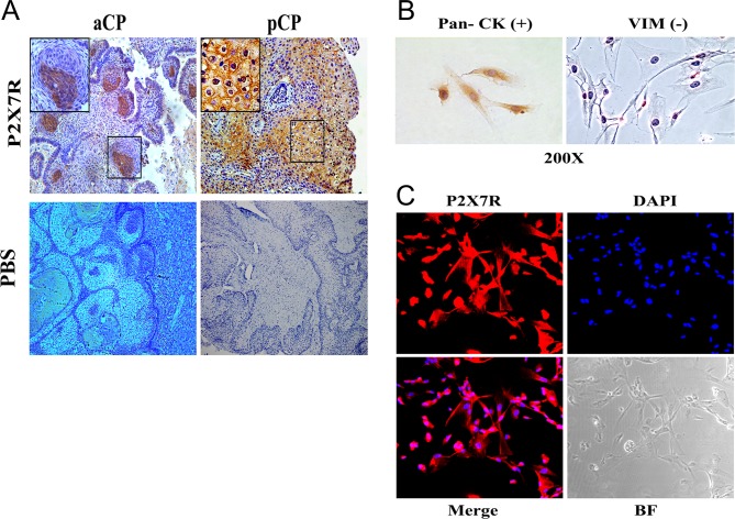
Figure 2.
Expression of the purinergic P2X7R in vivo and in cultured cells. immunohistochenistry of a tissue section of an adamantinomatous CP tumor (aCP, A) and a papillary CP tumor (pCP, B). Note labeling in whirl-like cellular structures in aCP and homogeneous staining in pCP. (C) Identification of primary aCP cells in culture. Tumor cells are characterized by positive staining for pan cytokeratin (pan CK, left panel) and absence of staining for vimentin (VIM, right panel). (D) Immunofluorescence for P2X7R along with DAPI labeling, merged panel and bright field (BF) microscopy as indicated. Note positive staining for P2X7R in tumor cells.

 This work is licensed under a