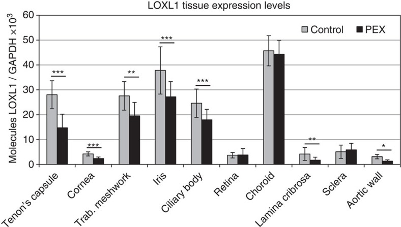
Figure 4. Determination of pathophysiologically relevant tissues and cell types.
Expression of LOXL1 mRNA in ocular and extraocular tissues derived from normal human donors (control) and donors with manifest PEX syndrome using real-time PCR technology. The expression levels were normalized relative to GAPDH. Expression levels were significantly reduced in Tenon's capsule (n=5), cornea (n=6), trabecular meshwork (n=17), iris (n=32), ciliary body (n=32), lamina cribrosa (n=20) and aortic wall (n=5) from PEX patients compared to controls, and were not different in retina, choroid and sclera (n=20 each); (data represent mean values±s.d.; *P<0.05, **P<0.001, ***P<0.0001; unpaired two-tailed Student's t-test).

