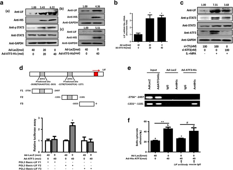
Fig. 4.

ATF3 transcriptionally increases LIF expression. (A, B) Ishikawa cells (a), primary endometrial epithelial cells (b) and stromal cells (c) were transduced with Ad-LacZ and Ad-ATF3-His at 0, 20 and 40 MOI for 48 h. (C) Ishikawa cells were exposed to E2 and MPA for 24 h after 48 h transfection with 0, 50, or 100 nM si-CTL or si-ATF3. LIF, STAT3, p-STAT3, HIS and GAPDH protein levels were measured by Western blotting assays. LIF mRNA expression levels were measured by qRT-PCR. *p < 0.05 vs. the Ad-LacZ group. The error bars indicate ± SD of 3 independent experiments. The density of LIF protein was analyzed and is presented. (D) Ishikawa cells were transfected with 500 ng firefly luciferase reporter plasmids (LIF-Luc) after transfecting Ad-His-ATF3 or Ad-LacZ for 24 h. Luciferase assays were performed, and the resulting data were normalized to constitutive Renilla luciferase levels (n = 3). (*p < 0.05). (E) Co-precipitated chromatin was amplified by PCR using primers specific for the LIF promoter region. PCR products were separated by agarose gel electrophoresis. Input (non-precipitated) chromatin was used as a positive control for these analyses. (F) Antibodies against LIF or mouse preimmune IgG were cultured with treated cells at a concentration of 0.5 μg/mL for 1 h before the transfer of the BeWo spheroids. **p < 0.01 Ad-ATF3 (b) vs. Ad-LacZ (a); # p < 0.05 anti-ATF3 (d) vs. IgG control (d). The error bars indicate ± SD of 3 independent experiments
