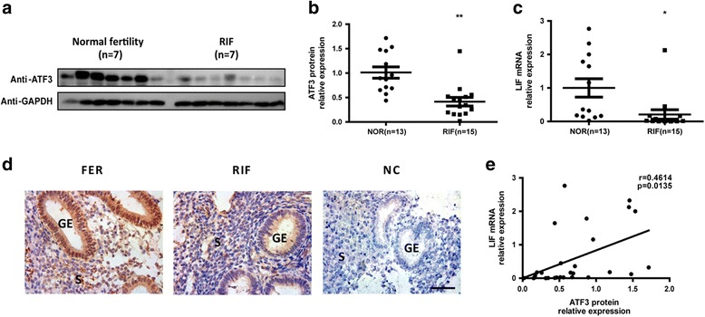
Fig. 5.

Aberrantly low ATF3 expression in the endometria of RIF patients. a Timed midsecretory endometrial biopsies from women with fertilized embryos (FER) (control, n = 7) and repeated implantation failure (RIF) patients (n = 7) were analyzed for ATF3 protein expression using Western blot analysis. Biopsies from FER women (control, n = 13) and RIF patients (n = 15) were analyzed for ATF3 protein expression by Western blot analysis (normalized to the GAPDH protein expression level) (b) and LIF mRNA level by real-time PCR (c). *p < 0.05; **p < 0.01 vs. the control group. d Secretory endometrial tissue samples from fertile control and RIF patients are shown at 400× magnification. The negative control (NC) is nonspecific rabbit serum. Brown staining represents positive staining (arrows). Scale bars, 50 μm. e Correlation between ATF3 protein expression and LIF mRNA expression in endometrial samples of FER women and RIF patients
