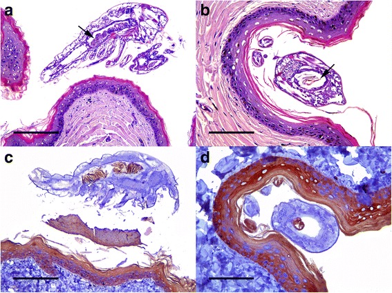
Fig. 4.

a Skin of a flipper. Parasite situated over the epidermis showing abundant granulated brownish material into its gut (arrow). Hematoxylin and eosin; scale bar = 200 μm. b Skin of a flipper. A parasite attached to the keratinized layer of the epidermis. The transversal section of the parasite allows observing brown laminated material that resembles keratin and small black spots that resemble melanin granules in gut content (arrow). Hematoxylin and eosin; scale bar = 100 μm. c Longitudinal and d cross sections of a parasite. Gut content reacts positively for cytokeratin. Anti-cytokeratin monoclonal antibody and ABC; scale bars = 200 and 100 μm, respectively
