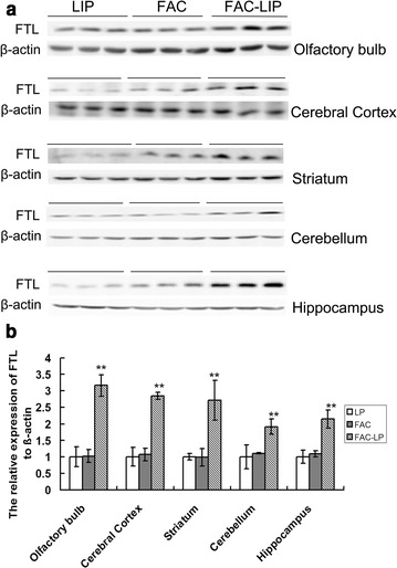
Fig. 4.

FTL protein expression in rat brain after intranasal administration of LIP, FAC and FAC-LIP. The olfactory bulb, cerebral cortex, striatum, cerebellum and hippocampus were dissected and the expression of FTL protein was detected with western blotting as described in “Methods”. a Shows the immunostained bands for FTL (21kD) and β-actin (42 kD), the density of the bands was analyzed and the statistical data was presented in b. Relative expression levels were normalized by β-actin and expressed as the mean ± SD. **P < 0.01 vs. LIP group. n = 6
