Fig. 7.

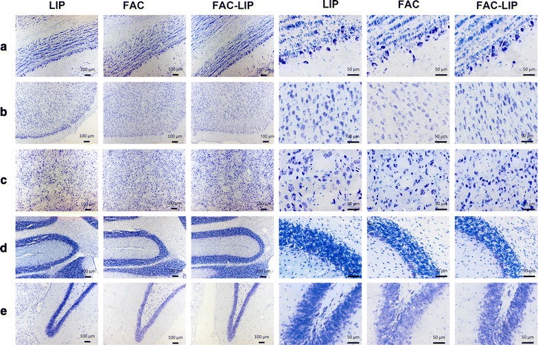
Cell morphology of brain tissues using Nissl staining after transnasal administration of LIP, FAC and FAC-LIP. a olfactory bulb, b cerebral cortex, c striatum, d cerebellum, e hippocampus, n = 3

Cell morphology of brain tissues using Nissl staining after transnasal administration of LIP, FAC and FAC-LIP. a olfactory bulb, b cerebral cortex, c striatum, d cerebellum, e hippocampus, n = 3