Fig. 2.
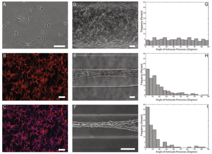
Astrocytes show starkly different morphology and alignment on 2-D surfaces and hydrogels of decreasing diameter. Astrocytes grown on a 2-D surface were found to show stellate morphology, whereas astrocytes grown in hydrogel micro-columns were found to increase alignment as a function of decreasing inner diameter. Phase contrast microscopy revealed that (A) astrocytes grown on a 2-D surface displayed characteristic stellate morphology. Confocal microscopy confirmed astrocytic phenotype following (B, C) immunostaining for astrocyte somata/processes (GFAP; red) and (C) Hoechst nuclear counterstain (blue). Further, astrocytes were seeded at high density in micro-columns with IDs of (D) 1.0 mm, (E) 350 μm, or (F) 180 μm. Astrocytes in (G) 1.0 mm ID micro-columns (n = 259 astrocytes from N = 6 micro-columns) did not exhibit a preferential alignment, whereas astrocytes cultured in (H) 350 μm ID (n = 428; N = 10) or (I) 180 μm ID (n = 114; N = 5) were induced to form an aligned morphology (p < 0.05 for each), as measured based on the percentage of processes growing at discrete angles with respect to the long axis of the micro-column (defined as 0°). This suggests that the physical restriction and radius of curvature of the micro-columns dictated the direction of astrocyte process outgrowth and morphology. Scale bars: 100 μm. (For interpretation of the references to colour in this figure legend, the reader is referred to the web version of this article.)

