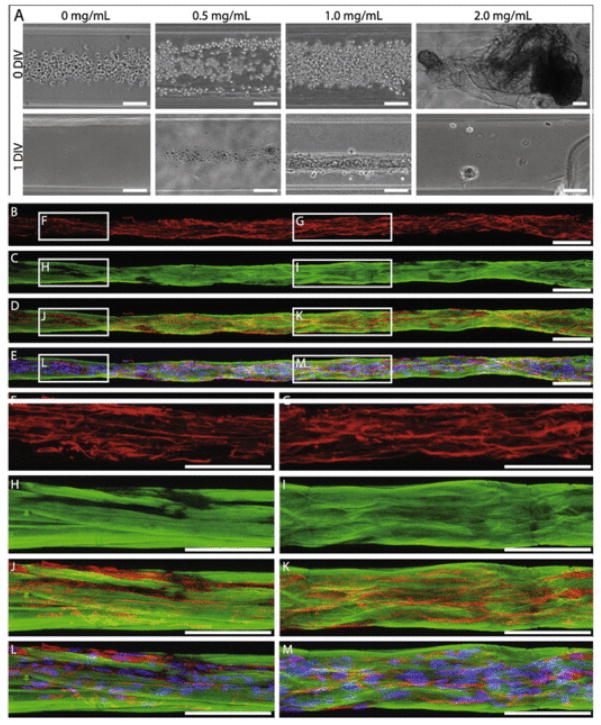
Fig. 4.
Collagen I concentration of 1.0 mg/mL uniquely supported astrocyte bundle formation. (A) Bright-field microscopy revealed that astrocytes seeded at high density in 350 μm ID micro-columns coated with 1.0 mg/mL collagen I adhered and formed dense longitudinal bundles over 1DIV, whereas astrocytes seeded in micro-columns with 0.0, 0.5, or 2.0 mg/mL did not adhere or exhibit growth at 1 DIV (N = 5 for each concentration). Note that collagen at 2 mg/mL was extruded from the micro-column interior. (B–M) Representative confocal reconstructions of the dense astrocyte-collagen bundles grown in 1.0 mg/mL collagen I stained via immunocytochemistry to denote (B, D–G, J–M) astrocyte somata/processes (GFAP; red) with (E, L, M) Hoechst nuclear counterstain (blue) and (C–E, H–M) collagen (collagen I; green). This confirmed that the longitudinal bundles were comprised of astrocytic somata/processes surrounded by a matrix and sheath of collagen. Scale bars: 100 μm. (For interpretation of the references to colour in this figure legend, the reader is referred to the web version of this article.)

