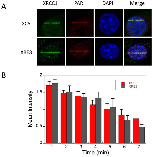
Fig. 1.
Immunofluorescent imaging of XRCC1 and PAR in XC5 and XRE8 cells. A) Cells were micro-irradiated in stripes to initiate XRCC1 recruitment and PAR synthesis and were fixed after 1 min repair time. Immunofluorescent staining methods (anti-XRCC1 antibody with Alexa 488 conjugated anti-mouse secondary antibody and anti-PAR antibody with Alexa 647 conjugated anti-chicken secondary antibody) are outlined in Section 2.2. Representative cells are shown. B) Time course of recruitment to damaged sites of endogenous XRCC1 in wild-type complemented XC5 cells and C12A reduced XRCC1 in XRE8 cells after fixing cells at the repair times specified and staining as outlined above. At least 5 cells were analyzed at each time point, error bars represent SEM.

