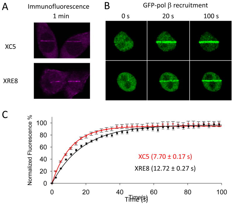
Fig. 2.
Pol β recruitment to UV micro-irradiation damage sites. (A) XC5 and XRE8 cells were irradiated in stripes and endogenous pol β recruitment was analyzed by immunofluorescence (anti-pol β antibody with Alexa 546 conjugated ant-rabbit secondary antibody) after 1 min. (B) Recruitment of transiently expressed GFP-pol β (see section 2.2.) was followed for at least 3 min after DNA damage. Images of representative cells after recruitment for 20 and 100 s are shown. (C) Plot of time course (100 s) of recruitment of GFP-pol β in XC5 and XRE8 cells. Experiments and data analysis are described in Section 2.2. Fluorescence data were normalized using intensity at the beginning of recruitment and maximal intensity values and fitted to a single exponential. Nineteen cells were analyzed in each case, and error bars represent SEM. The half-time of pol β recruitment in XC5 cells (7.70 ± 0.17 s) was statistically different (p<0.001) to the that in XRE8 cells (12.72 ± 0.27 s).

