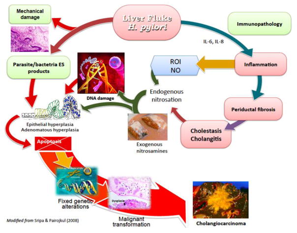
Figure 3. Hypothesized pathways of pathogenesis of opisthorchiasis/H. pylori-induced cholangiocarcinoma.
The liver fluke Opisthorchis viverrini/Helicobacter damages bile duct tissue via at least three distinct pathways: 1) mechanical damage to biliary epithelia caused by parasites sucking; 2) inflammation-induced immunopathology, particularly due to reactive oxygen intermediates (ROI) and nitric oxide (NO); and 3) direct effects of fluke/Helicobacter secreted proteins on biliary epithelia including cell proliferation induced by parasite-derived growth factors. These pathways converge, resulting in genetic lesions and unregulated proliferation. Damaged DNA/genes after successive replications become fixed, leading to malignant transformation of cholangiocytes. Adapted from [32].

