Fig. 8.

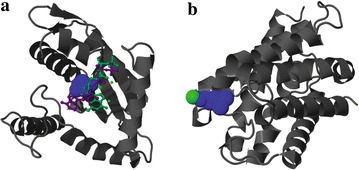
Three-dimensional (3D) models of a. protein 5 and b. protein 6. Background helices shown in gray, Active sites shown in blue, Hypothetical ligands shown in magenta and green

Three-dimensional (3D) models of a. protein 5 and b. protein 6. Background helices shown in gray, Active sites shown in blue, Hypothetical ligands shown in magenta and green