Figure 4.

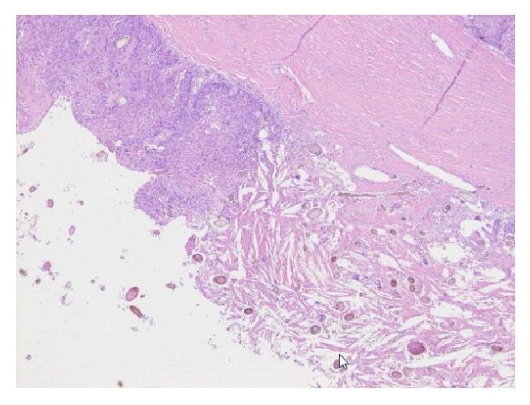
Histologically, squamous cell carcinoma was observed inside the cyst (left side of the picture). Granulation tissue containing hair was also found (right side of the picture) (H&E staining, ×100).

Histologically, squamous cell carcinoma was observed inside the cyst (left side of the picture). Granulation tissue containing hair was also found (right side of the picture) (H&E staining, ×100).