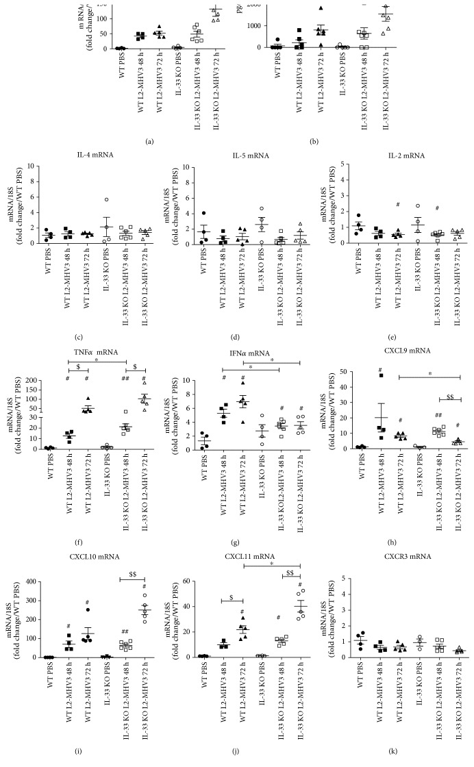
Figure 7.
Expression of TH1/TH2 cytokines and chemokines in WT and IL-33 KO mice during L2-MHV3 hepatitis. Total liver RNA was extracted from WT or IL-33 KO mice and control (PBS) or L2-MHV3-infected mice (48 h and 72 h PI) and tested by RT-qPCR for (a) IL-6, (c) IL-4, (d) IL-5, (e) IL-2, (f) TNFα, (g) IFNγ, (h) CXCL9, (i) CXCL10, (j) CXCL11, and (k) CXCR3. The mean expression of the WT PBS mice was used as a control and arbitrarily considered as 1 AU (arbitrary unit) which served as a reference for fold change in other conditions. (b) Sera from WT or IL-33 KO mice (PBS) or L2-MHV3-infected mice (48 h and 72 h PI) were quantified for IL-6 expression. Statistical analyses were done according to the Mann-Whitney U test. # represents significant difference with WT PBS mice, ∗ represents significant difference between WT and IL-33 KO mice, and $ represents significant difference between two conditions in the same background mice. #p < 0.05; ##p < 0.01; and ###p < 0.001.

