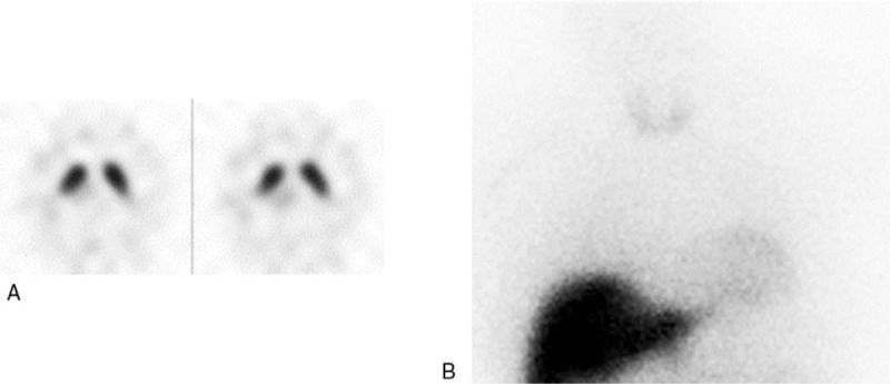
Figure 3.

A 55-year-old male patient with uncertain parkinsonism, vascular lesions in left basal ganglia and midbrain at MRI, and partial response to dopamine agonist treatment. 123I-Ioflupane SPECT was normal (A) with also BP values above cut-off in caudate and putamen nuclei, bilaterally; 123I-MIBG cardiac scintigraphy was pathological (B) with reduced H/M values both in early (1.43) and delayed (1.38) phases. The patient was finally confirmed with uncertain parkinsonism and monitored in a close follow up. H/M = heart to mediastinum ratio, MRI= magnetic resonance imaging, MIBG = metaiodobenzylguanidine, SPECT = single photon emission computed tomography.
