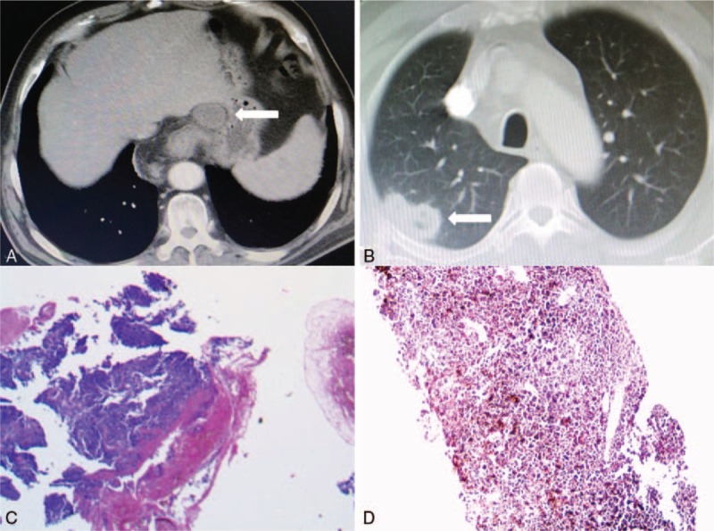
Figure 1.

(A) Computed tomography (CT) scan on admission showed a tumor measuring 5.5 cm × 3.5 cm × 3.0 cm in the lower esophagus with enlarged celiac lymph nodes (straight arrow). (B) The concurrent pulmonary lesion of 2.0 cm × 1.0 cm in size located in right upper lobe, (C, D) Postoperative histopathology revealed esophageal and pulmonary melanoma, by H&E staining (×100).
