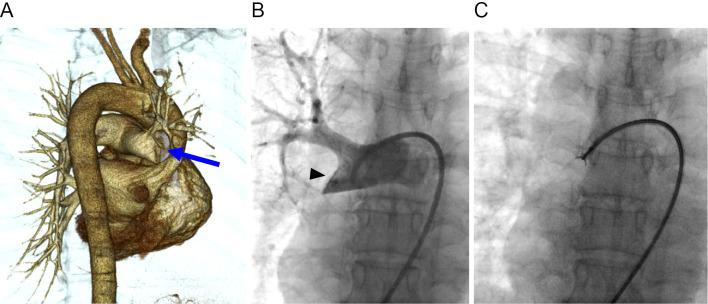
Figure 2.
Angiographic examinations. (A) Three-dimensional computed tomographic angiography (posterior oblique view) shows complete occlusion of the right distal main pulmonary artery (arrow). (B) pulmonary angiography shows a filling defect occupying the entire lumen of the right distal main pulmonary artery (arrowhead). (C) An endovascular catheter biopsy using forceps is performed following pulmonary angiography.

