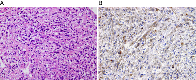
Figure 3.
Histological examinations. (A) The tumor is composed of fascicular and poorly-arranged proliferation of atypical spindle cells. The tumor cells are anaplastic, with atypical nuclei and some multinucleated cells (Hematoxylin and Eosin staining, original magnification 200×). (B) The tumor cells are immunohistochemically positive for platelet-derived growth factor receptor α (original magnification 200×).

