. 2017 May 25;53(2):133–139. doi: 10.4068/cmj.2017.53.2.133

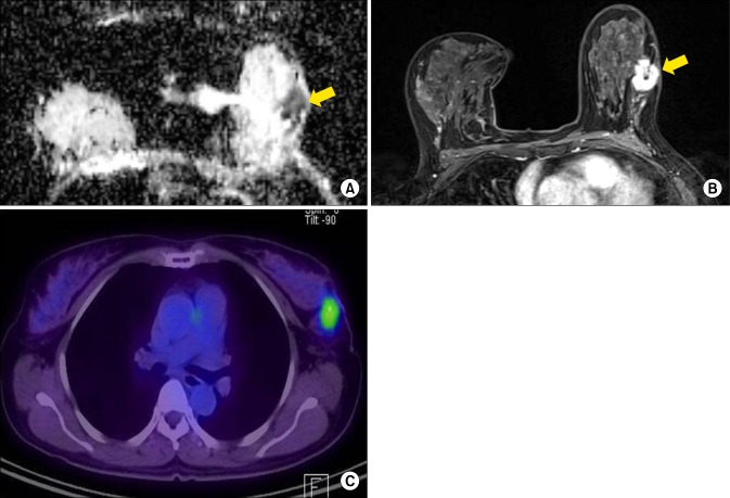
FIG. 1. A 64-year-old woman with invasive lobular carcinoma of the left breast. (A) ADC map shows RIO for measuring the mean ADC value. The calculated ADC value was 1,115×10−6 mm2/s, which was slightly higher than mean ADC value (1,117×10−6 mm2/s). (B) Axial enhanced T1-weighted image after two minutes of contrast injection demonstrates an irregular heterogeneous enhancing mass in the left breast. Background parenchymal enhancement was grade 3, moderate enhancement. (C) FDG PET/CT image shows FDG uptake in the left breast with measured SUVmax as 3.7. On histologic examination, axillary LN metastasis was noted in two LNs. The results of immunohistochemical study were ER (+), PR (+), HER2 (−), EGFR (+) and Ki-67 2%. ADC: apparent diffusion coefficient, EGFR: epidermal growth factor receptor, ER: estrogen receptor, FDG PET/CT: 18 F-fluorodeoxygluxose positron emission tomography/computed tomography, HER2: human epidermal growth factor receptor 2, EGFR: epidermal growth factor receptor, LN: lymph node, PR: progesterone receptor, SUVmax: maximum standardized uptake value.

FIG. 1