. 2017 May 25;53(2):133–139. doi: 10.4068/cmj.2017.53.2.133

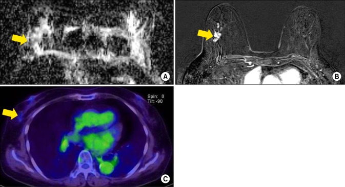
FIG. 2. A 71-year-old woman with invasive lobular carcinoma of the right breast. (A) On ADC map, calculated ADC value by ROI was 1,260×10−6 mm2/s, which was higher than mean ADC value (1,117×10−6 mm2/s). (B) Axial enhanced T1-weighted image shows nonmass like enhancement in the right breast. BPE was grade 1, minimal enhancement. (C) FDG PET/CT image shows minimal FDG uptake in the right breast with measured SUVmas as 1.3. On histologic examination, there was no axillary LN metastasis. Immunohistochemical study showed ER (+), PR (−), HER2 (−), EGFR (+) and Ki-67 10%. ADC: apparent diffusion coefficient, EGFR: epidermal growth factor receptor, ER: estrogen receptor, FDG PET/CT: 18 F-fluorodeoxygluxose positron emission tomography/computed tomography, HER2: human epidermal growth factor receptor 2, EGFR: epidermal growth factor receptor, LN: lymph node, PR: progesterone receptor, SUVmax: maximum standardized uptake value.

FIG. 2