. 2017 May 25;53(2):83–94. doi: 10.4068/cmj.2017.53.2.83

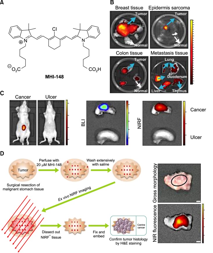
FIG. 3. Preferential uptake and retention of MHI-148. (A) MHI-148 chemical structure. (B) Ex vivo NIR fluorescence imaging showed increased MHI-148 dye uptake by different types of canine spontaneous tumors (blue arrows) as compared to adjacent normal tissues (white arrows). (C) Preferential uptake of NIRF dye in gastric cancer tissues relative to that in gastric tissues. In vivo NIRF imaging of mice bearing either orthotopic luc-tagged gastric tumor xenografts (left) or gastric ulcers (right). (D) NIRF imaging of clinical gastric cancer tissues. Schematic outlining the experimental procedures for NIRF imaging of freshly resected clinical gastric tumor tissues (left). Gross morphology and NIRF imaging of representative tumor tissues surgically resected from one of three gastric cancer patients. Representative images are presented in all panels. Original magnification: 4×; scale bars represent 4 mm. Figure images and the accompanying legend are reproduced with permission from (25,30). Copyright © 2017 Impact Journals.

FIG. 3