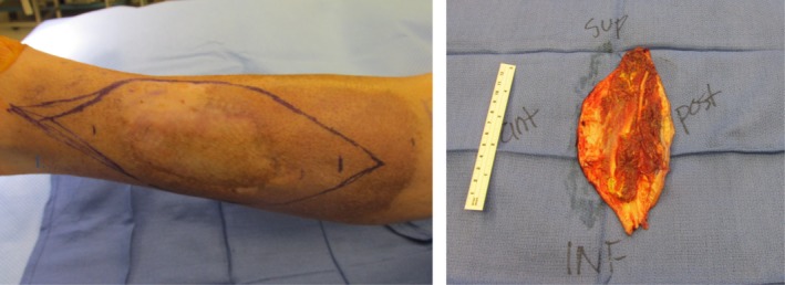
Figure 3.

Preoperative photograph showing a central area of residual sarcoma, surrounding hyperpigmentation following external beam radiation therapy, and demarcation of planned surgical incision for adequate tumor margin. Postoperative photograph showing the orientation and posterior depth of invasion of the surgical specimen following wide local excision.
