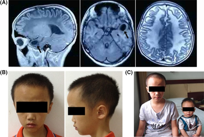
Figure 1.

(A) Brain MRI of the proband. The image shows a subcortical cyst in the left temporal lobe (arrow) and a diffuse lesion in the white matter. (B) The patient's photographs at the age of nine. The head circumference was large. (C) Proband (left) and his healthy brother (right). The baby was about ten months old. No abnormity was found.
