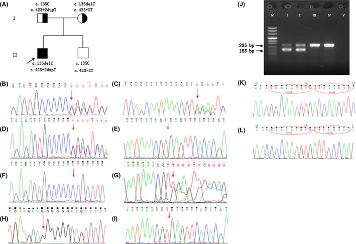
Figure 2.

Genetic analysis. (A) Pedigree chart. The proband was noted with compound heterozygous mutations. The father and mother both carried heterozygous mutations. The prenatally diagnosed boy was free of mutations. (B, C) Sequence of the proband's sample. A: Arrow pointing to the heterozygous mutation c.135delC; B: heterozygous mutation c.423+2dupT. (D, E) Sequences of the two mutation regions in the mother. (F, G) Sequences of the two mutation regions in the father. (H, I) gDNA sequence of amniotic fluid. H: Arrow pointing to the normal c.135C; I: arrow showing the position of normal c.423+2T. (J) Electrophoretogram of cDNA amplification. “M” is the DNA marker; “I”, “II”, “III”, “IV,” and “V” indicate the proband, the father, the mother, normal, and blank control, respectively. There were two separable bands in the samples of the proband and the father. The top band was 285 bp in size; the lower one was 183 bp. The mother's RT‐PCR product was equal to the normal control. (K, L) cDNA sequence of the proband. K: The smaller product was 183 bp, and red ovals at the bases of MLC1 mark exon 4 and exon 6. L: Product of normal length, the bases in the red ovals belong to exon 4 and exon 5 of MLC1.
