
Figure 2.
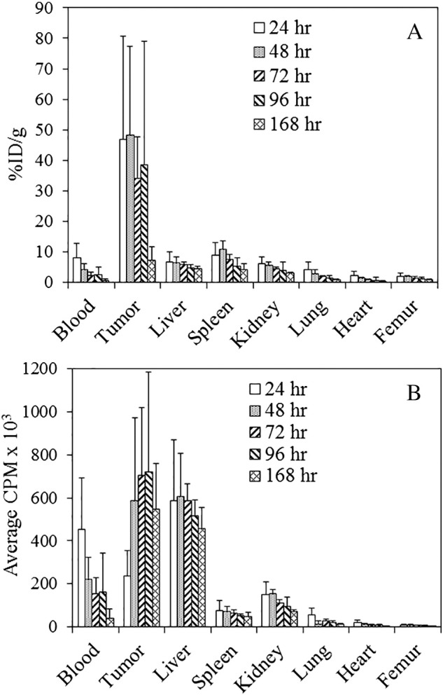
Tumor and normal tissue distribution of 111In-panitumumab.
(A) Athymic mice bearing 5-day LS-174T i.p. tumor xenografts were injected i.p. with 111In-panitumumab (~7.5 μCi) and euthanized (n = 5) at 24, 48, 72, 96, and 168 hours after the injection. The tumor and tissues were harvested and wet-weighed, and the radioactivity was measured in a γ-counter. The %ID/g and standard deviation were calculated. (B) The average cpm of the tumor and tissues was also plotted along with the standard deviation.