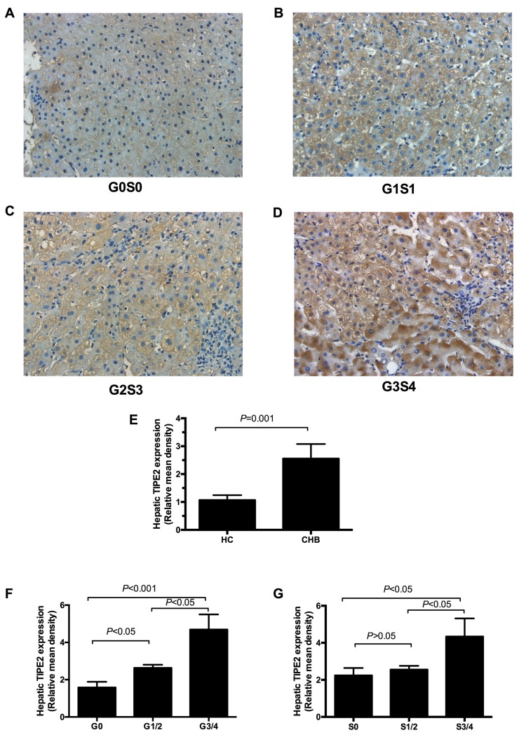
Figure 3. Expression of TIPE2 protein in liver tissue of CHB patients.
A.-D. The positive staining for TIPE2 was found mainly in the hepatocytes (200×). E. Relative mean density analysis showed the difference in hepatic TIPE2 staining between CHB group and healthy controls. F. Expression of hepatic TIPE2 staining between inflammation grade 0, grade (1-2) and grade (3-4). G. Expression of hepatic TIPE2 staining between fibrosis stage (3-4), stage (1-2), and fibrosis stage 0.

