
Figure 7. Induction of ER stress leads to 2-DG induced cell death and GC sensitization.
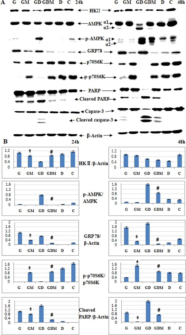
(A) Western blot analysis of HKII, AMPKα, p-AMPKα (Thr172), GRP78, p70S6K, p-p70S6K (Thr421/Ser424) and cell apoptosis associated proteins in Molt-4 cells after 48 h exposure to 2-DG, Dex, mannose, alone or in combination. β-Actin was used as an internal control. (B) Bar graphs show the ratio of protein to β-Actin and phospho-protein to total protein. For all experiments, values of triplicate experiments are shown as the mean ± SD. *p < 0.01 versus the 2-DG group. #p < 0.01 versus the 2-DG+Dex group. G, 2-DG group; GM, 2-DG+mannose group; GD, 2-DG+Dex group; GDM, 2-DG+Dex+mannose group; D, Dex group and C, control group.