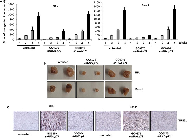
Figure 7. Induction of apoptosis in the xenografted tumors.

(A) Cells were inoculated subcutaneously into the nude mice. GO6976 was injected peritoneally after the inoculation and subsequently administrated every 3 days. One week later, the diameters of the tumors were measured and then every week for consecutive 4 weeks. The error bars represent the SD (n = 4, *p < 0.05). (B) After the tumors were dissected from the mice, the pictures of the tumors were taken. (C) The slides mounted with the tumor samples were stained with TUNEL reagent.
