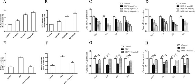
Figure 5. DBP generates oxidative stress and reduces the expression of the gene responsible for the prevention of oxidative activity in NRK49F and NRK52E cells.

(A) Measurement of intracellular ROS in NRK49F cells by DCFH-DA dye showed the quantity of ROS increased significantly after treatment with various concentrations of DBP (1, 10, 100 μmol/L). (B) Measurement of intracellular ROS in NRK52E cells by DCFH-DA dye showed the quantity of ROS increased significantly after treatment with various concentrations of DBP (1, 10, 100 μmol/L). (C) Real-time PCR assessed the expression of antioxidant genes (Gpx1, Cat, GR, Gst) of NRK49F cells exposed to various concentrations of DBP (1, 10, 100 μmol/L). (D) Real-time PCR assessed the expression of antioxidant genes (Gpx1, Cat, GR, Gst) of NRK52E cells exposed to various concentrations of DBP (1, 10, 100 μmol/L). (E) Measurement of intracellular ROS by DCFH-DA dye in NRK49F cells in DBP-exposed, vitamin C-exposed group and unexposed controls. (F) Measurement of intracellular ROS by DCFH-DA dye in NRK52E cells in DBP-exposed, vitamin C-exposed group and unexposed controls. (G) Real-time PCR assessed the expression of antioxidant genes (Gpx1, Cat, GR, Gst) of NRK49F cells in DBP-exposed, vitamin C-exposed group and unexposed controls. (H) Real-time PCR assessed the expression of antioxidant genes (Gpx1, Cat, GR, Gst) of NRK52E cells in DBP-exposed, vitamin C-exposed group and unexposed controls. Data represent mean ± SD. n = 6 rats per group, *P < 0.05.
