
Figure 5.
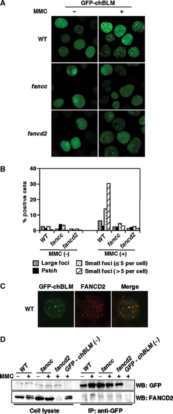
Analysis of GFP-chBLM in wild-type, fancc, and fancd2 DT40 cells. (A) Focus formation of GFP-chBLM. Cells expressing GFP-chBLM were observed before and 7 h after MMC treatment (500 ng/ml, 1 h). (B) Fraction of cells having large foci, indicated number of small foci, and patch-like accumulation. Cells were treated as in (A). At least 200 cells were scored in each preparation. (C) Colocalization of GFP-chBLM and FANCD2 in wild-type cells treated with MMC as in (A). (D) Co-immunoprecipitation of GFP-chBLM with FANCD2. Cells with indicated genotypes were treated with MMC (500 ng/ml for 7 h) or left untreated and lysed. A portion of the lysate (2.5%) was saved as an input control, and the rest was subjected to immunoprecipitation with anti-GFP. The lysates and immunoprecipitates were separated and probed with the indicated antibodies. GFP-chBLM (−), wild-type cells that were not transfected with the GFP-chBLM construct serving as negative control. The experiments were repeated three times with similar results.