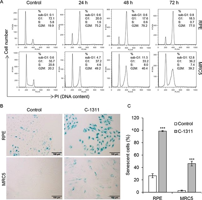
Figure 5. Increased senescence in non-cancer cells treated with C-1311.

(A) Retinal pigment epithelial RPE cells and fetal lung fibroblast MRC-5 cells were treated with C-1311 (0.68 μM) for the times indicated and DNA content was determined following PI staining by FACS. Histograms are representative of three independent experiments. (B and C) RPE and MRC5 cells were exposed to C-1311 (0.68 μM) for 120 h, stained for SA-β-gal activity. (B) Representative images from bright-field microscope. (C) Quantitation of the percentage of senescent cells. The data are presented as mean ± SD, n = 3. ***P < 0.001 vs control group.
