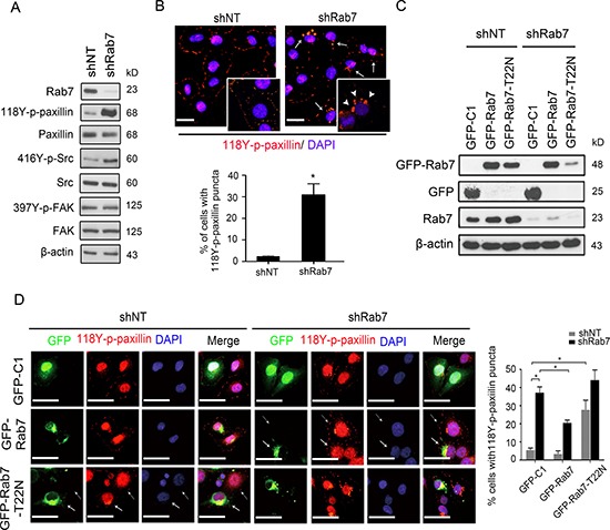
Figure 2. Rab7 and its GTPase activity are required for paxillin accumulation into cytosolic puncta.

(A) Lysates from shNT or shRab7-expressing BT-20 cells were immunoblotted with antibodies against Rab7, LC3B, 118Y-p-paxillin, paxillin, 397Y-p-FAK, FAK416Y-p-Src, Src and β-actin as an internal control. (B) Top: shNT and shRab7 transfected BT-20 cells were fixed and stained with anti-118Y-p-paxillin antibody (red) and with DAPI (blue). Solid arrows identify cells with 118Y-p-paxillin puncta. Arrowheads in inserts (65x magnification) indicate actual 118Y-p-paxillin puncta. Bottom: percentage of cells with 118Y-p-paxillin puncta. Data are presented as mean ± SEM (*p < 0.05, n = 3). (C and D) BT-20 cells expressing shNT and shRab7 plasmids and their matched cells rescued with empty (GFP-C1), shRNA-resistant Rab7 (GFP-Rab7) or Rab7 with a point mutation (GFP-Rab7-T22N) plasmids, were lysed and immunoblotted with anti-GFP antibody (C) or were fixed and stained with anti-118Y-p-paxillin antibody (red) and with DAPI (blue). Scale bar, 20 μm. Solid arrows indicate the cell expressing GFP plasmids and dashed arrows indicate cells without expressing GFP plasmids (D, left). (D, right) Graph shows the quantification of percentage of cells with 118Y-p-paxillin in intracellular puncta (determined using lower magnification images (20 ×)). Data are presented as mean ± SEM (*P < 0.05, n = 3)
