
Figure 6. Tyrosine 118 phosphorylation of paxillin modulates its association with the autophagic marker LC3 at cell plasma membrane protrusions.
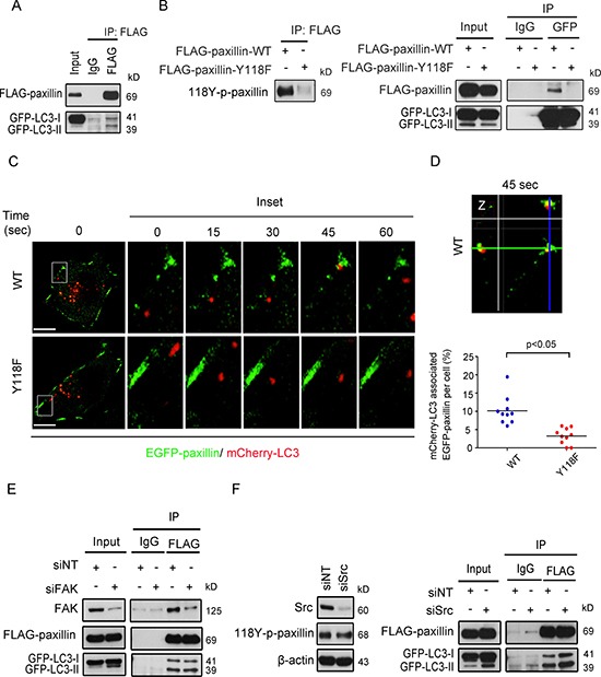
(A) BT-20 cells were transfected with FLAG-paxillin and GFP-LC3 plasmids for 24 h and immunoprecipitated with either anti-IgG or anti-FLAG antibodies. Lysates were then immunoblotted with anti-FLAG and anti-GFP antibodies. (B) Left: FLAG protein was immunoprecipitated from BT-20 cells transfected with either wild-type FLAG-paxillin (FLAG-paxillin-WT) or FLAG-paxillin with point mutation at amino acid 118 (FLAG-paxillin-Y118F) for 24 hours and immunoblotted with anti-118Y-p-paxillin antibody. Right: BT-20 cells were transfected with either FLAG-paxillin-WT or FLAG-paxillin-Y118F and GFP-LC3 plasmids for 24 h and immunoprecipitated with either anti-IgG or anti-GFP antibodies. Lysates were then immunoblotted with anti-FLAG and anti-GFP antibodies. (C) BT-20 cells were transfected with either EGFP-paxillin wild-type (WT) or EGFP-paxillin-Y118F (Y118F) plasmids (green) and mCherry-LC3 plasmids (red) for 24 h, then plated on chamber slide. Next, cells were monitored for the trafficking of EGFP-paxillin and mCherry-LC3 by time-lapse spinning disc microscopy. Scale bar, 5 μm. Higher-magnification images of the inserts are also shown indicating path of EGFP-paxillin and mCherry-LC3 at cell protrusions. (D) Top, Enlargements and cross-sections of the confocal-z-planes of the boxed regions are also shown indicating association of EGFP-paxillin-WT and mCherry-LC3 at 45 sec. Bottom: quantification of the percentage of dynamic EGFP-paxillin labeling FAs per cell targeted by mCherry-LC3. Scatter plot shows individual single cells and mean line. n = 125 mCherry-LC3 vesicles targeted to FA in EGFP-paxillin WT and 169 mCherry-LC3 vesicles targeted to FAs in EGFP-paxillin-Y118F group from 10 single cells. (E) siRNA silenced FAK BT-20 cells were transfected with FLAG-paxillin and GFP-LC3 plasmids for 24 h and immunoprecipitated by either anti-IgG or anti-FLAG antibodies. Lysates were then immunoblotted with anti-FAK, anti-FLAG and anti-GFP antibodies. (F) Left: BT-20 cells were transiently transfected with non-targeted or Src siRNA proteins were extracted, and then immunoblotted with anti-Src, anti-118Y-p-paxillin and anti-β-actin antibodies. Right: FLAG-paxillin was immunoprecipitated from siNT and siSrc cells expressing FLAG-paxillin and GFP-LC3, then immunoblotted with anti-FLAG and anti-GFP antibodies.