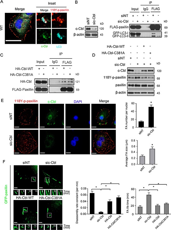
Figure 7. c-Cbl is the major cargo receptor mediating LC3 interaction with paxillin.

(A) BT-20 cells were fixed and stained for anti-118Y-p-paxillin antibody (red), anti-LC3B antibody (cyan), anti-c-Cbl antibody (green) and with DAPI (blue). Scale bar, 20 μm. Enlargements of the boxed regions are also shown indicating association of 118Y-p-paxillin, LC3B and c-Cbl. (B) Left: BT-20 cells were transfected with either NT or c-Cbl siRNA for 48 h, then immunoblotted with anti-c-Cbl anti-β-actin antibodies. Right: BT-20 cells were transfected with non-targeted or c-Cbl siRNA for 24 h and then transfected with FLAG-paxillin and GFP-LC3 plasmids for another 24 h and immunoprecipitated by either anti-IgG or anti-FLAG antibodies. Lysates were then immunoblotted with anti-FLAG and anti-GFP antibodies. (C) BT-20 cells were transfected with either HA-Cbl wild-type plasmids (HA-Cbl-WT) or HA-Cbl with point mutation on a.a 381. (HA-Cbl-C381A) for 24 h and immunoprecipitated by either anti-IgG or anti-FLAG antibodies. Lysates were then immunoblotted with anti-FLAG and anti-HA antibodies. (D) BT-20 cells were transfected with non-targeted or c-Cbl siRNA for 24 h and then were either rescued by HA-Cbl-WT or HA-Cbl-C381A plasmids. After 24 h, cells were lysed and immunoblotted with antibodies against c-Cbl, 118Y-p-paxillin, paxillin and anti- β-actin as an internal control. (E) Left, BT-20 cells transfected with non-targeted or c-Cbl siRNA for 48 h were stained with anti-118Y-p-paxillin antibody (red), c-Cbl antibody (green) and DAPI (blue). Scale bar, 20 μm. Right, quantification of FA number per cells and average FA size. Data are presented as mean ± SEM (*p < 0.05, n = 10 cells) (F) Left: Serum-starved siNT or sic-Cbl BT-20 cells rescued with either HA-Cbl wild-type plasmids (HA-Cbl-WT) or HA-Cbl with point mutation on a.a 381 (HA-Cbl-C381A) were transfected with GFP-paxillin for 24 h, then plated on chamber slide, stimulated by 20 ng/ml EGF and analyzed for FA turnover by time-lapse spinning disc microscopy. Scale bar, 2 μm. Higher-magnification images of the inserts are also shown indicating positions of paxillin-containing FAs. Right: FAs disassembly rate and FA life time in siNT, sic-Cbl. sic-Cbl+ HA-Cbl-WT and sic-Cbl+ HA-Cbl-C381A cells were quantified (mean ± SEM, n = 44 FAs in siNT, 38 FAs in sic-Cbl, 45 FAs in HA-Cbl rescued and 52 FAs in HA-Cbl-C381A groups from 10 single cells, *p < 0.05).
